

Key Takeaways
Option A
Tendons & Ligaments
The body's fibrous cables - connecting and stabilizing moving parts.
Best for: Understanding how muscles transmit force and how bones stay properly aligned during movement.
Option B
Cartilage
The body's resilient cushioning layer - smooth, flexible, and shock-absorbing.
Best for: Understanding how joints withstand decades of repetitive loading without bone grinding on bone.
If you want to understand why a muscle tear feels different from a joint sprain
Tendons & Ligaments
Tendons anchor muscles to bones while ligaments stabilize bone-to-bone connections - they are separate structures with distinct injury patterns and recovery timelines.
If you're trying to understand joint pain or conditions like osteoarthritis
Cartilage
Articular cartilage is the tissue that breaks down in osteoarthritis. Its lack of blood supply makes deterioration a major clinical challenge.
If you're curious about why connective tissue injuries tend to heal slowly
Tendons & Ligaments
While tendons have limited blood supply, ligaments have even less - both heal more slowly than muscle because collagen repair is a gradual, resource-limited process.
Three Tissues, Three Jobs
People often lump tendons, ligaments, and cartilage together as vague "soft tissue" - but each plays a distinct biological role. All three are forms of connective tissue, meaning they're built from cells embedded in a protein-rich matrix dominated by collagen. Beyond that shared origin, the similarities thin out quickly.
Tendons are dense, cord-like bands that anchor skeletal muscles to bone. When your bicep contracts, it's the tendon that transmits that pulling force across the elbow joint to move your forearm. Tendons are engineered for tensile strength - they resist stretching under load - and are arranged in tightly packed parallel collagen fibers to do exactly that. The Achilles tendon, connecting your calf muscles to your heel bone, is the largest and most recognizable example.
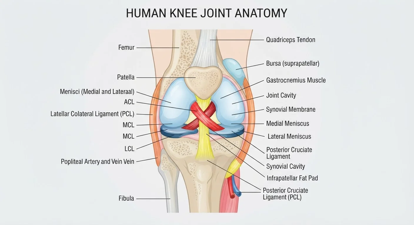
Ligaments do a different job: they connect bone to bone, primarily to constrain and stabilize joints. The anterior cruciate ligament (ACL) in the knee, for instance, prevents the tibia from sliding too far forward relative to the femur. Ligaments also contain collagen, but their fibers run in multiple directions rather than parallel - reflecting the multidirectional forces joints must resist. This architecture makes ligaments slightly more elastic than tendons, but still far less flexible than muscle.
To understand how these tissues fit into the broader skeletal system, see our article on why bones are living tissue, which covers the dynamic structure tendons and ligaments attach to.
What Cartilage Actually Does
Cartilage is structurally unlike either tendons or ligaments. Rather than transmitting force or stabilizing joints, it primarily functions as a low-friction, shock-absorbing surface. Cartilage is composed of specialized cells called chondrocytes suspended in a gel-like matrix rich in collagen and proteoglycans - large molecules that attract and retain water, giving cartilage its characteristic springiness.
There are three types of cartilage in the human body, each with a different composition suited to its location:
- Hyaline (articular) cartilage - the smooth, glassy tissue covering the ends of bones in joints like the knee and hip. It allows bones to glide past each other with minimal friction.
- Fibrocartilage - tougher and denser, found in intervertebral discs and the menisci of the knee. It handles compression and shear forces simultaneously.
- Elastic cartilage - flexible and resilient, found in the outer ear and epiglottis, where shape-retention under repeated bending matters.
Critically, cartilage is avascular - it has no direct blood supply. Nutrients diffuse in from surrounding joint fluid and nearby tissue. This is why cartilage heals so poorly after injury or wear: without blood vessels delivering repair cells, restoration is extremely limited. It's the central challenge behind conditions like osteoarthritis, in which articular cartilage progressively breaks down.
| Criterion | Tendons & Ligaments | Cartilage |
|---|---|---|
| Primary function | Connect and transmit force or stabilize joints | Cushion, reduce friction, absorb shock |
| What they connect | Muscle-to-bone (tendon) or bone-to-bone (ligament) | Covers bone ends; found in discs and airways |
| Main structural protein | Type I collagen (dense, parallel fibers) | Type II collagen + proteoglycans |
| Blood supply | Limited (tendons slightly more than ligaments) | None (avascular) |
| Healing capacity | Slow but possible with adequate rest | Very poor; significant damage often permanent |
| Common injury type | Strain (tendon) or sprain (ligament) | Wear, degeneration, or traumatic tear |
| Associated condition | Tendinopathy, ACL/MCL tears | Osteoarthritis, meniscus tears |
Injuries and Why They Matter
Understanding the distinction between these tissues has real practical value, particularly when it comes to injuries. A sprain refers specifically to a stretched or torn ligament - the kind of injury you get when you roll an ankle or take a bad hit to the knee. A strain, by contrast, involves a muscle or tendon. These terms are often misused interchangeably in everyday conversation, but clinically they indicate different structures and different treatment considerations.
Tendon injuries - such as Achilles tendinopathy or rotator cuff tears - often develop gradually from repetitive overuse rather than a single traumatic event. Ligament tears, like an ACL rupture, are more commonly acute. Both heal slowly due to limited blood supply, but tendons generally have slightly better vascularization than ligaments, giving them a marginal healing advantage.
Cartilage injuries are in a category of their own. Because chondrocytes don't regenerate efficiently after damage and blood-borne repair cells can't reach avascular cartilage, significant cartilage loss is often irreversible without surgical intervention. Emerging techniques such as autologous chondrocyte implantation (ACI) aim to address this limitation, though research is ongoing.
The musculoskeletal system doesn't operate in isolation either - for a broader look at how body systems coordinate, see body systems that work together. And if you're curious how muscles themselves generate the forces tendons transmit, our article on skeletal vs. smooth vs. cardiac muscle provides useful context.