

Key Takeaways
Skin as an Organ
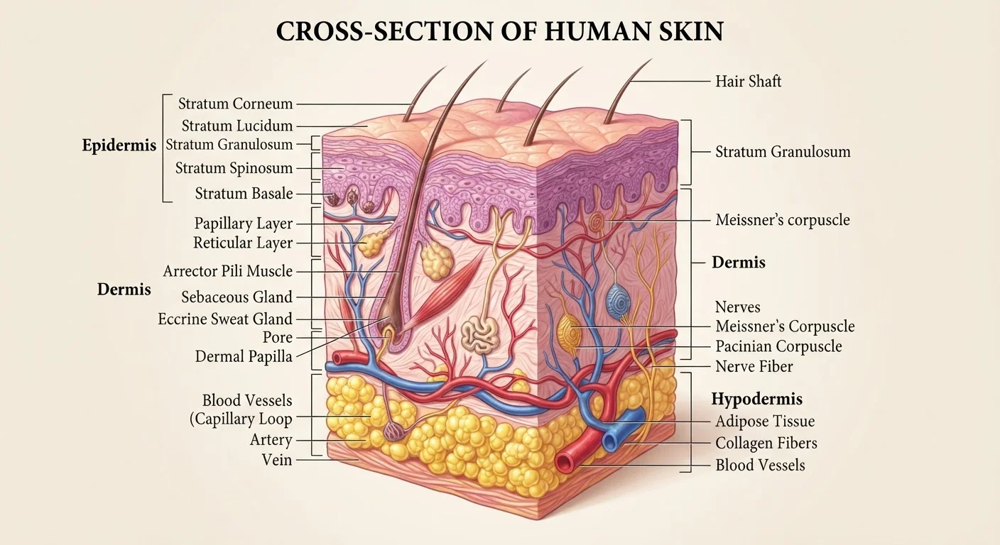
The skin is the human body's largest organ by surface area and weight. It consists of three distinct layers - the epidermis, dermis, and hypodermis - each performing specialized functions. Far from being a passive wrapper, skin actively regulates temperature, prevents infection, and communicates sensory information to the brain.
In adults, skin typically covers approximately 1.5 to 2 square meters of surface area and accounts for roughly 15% of total body weight, according to standard anatomical references.
The Epidermis: The Body's First Line of Defense
The epidermis is the outermost layer of skin - the part you can see and touch. It is composed primarily of cells called keratinocytes, which produce a tough structural protein called keratin. These cells are born at the base of the epidermis and gradually migrate upward, flattening and hardening before being shed from the surface in a continuous cycle known as keratinization.
The epidermis contains no blood vessels; it receives nutrients by diffusion from the dermis below. Specialized cells called melanocytes, scattered throughout its base layer, produce melanin - the pigment that determines skin color and absorbs ultraviolet (UV) radiation to protect deeper tissues from DNA damage. The epidermis also hosts Langerhans cells, immune sentinels that detect and respond to foreign pathogens, making the skin an active participant in immune surveillance.
The Dermis: Strength, Sensation, and Support
Directly beneath the epidermis lies the dermis, a thicker layer of connective tissue composed largely of collagen and elastin fibers. Collagen provides tensile strength, while elastin allows the skin to stretch and spring back - properties that decline with age and UV exposure, contributing to wrinkling and sagging.
The dermis is densely populated with functional structures. Sweat glands help regulate body temperature through evaporative cooling. Sebaceous glands secrete sebum, an oily substance that lubricates the skin and hair. Hair follicles anchor each strand of hair and cycle through phases of growth, rest, and shedding. Perhaps most importantly for daily experience, the dermis is rich with mechanoreceptors and nociceptors - specialized nerve endings that detect pressure, texture, vibration, temperature, and pain, feeding constant sensory data to the brain.
~2 m²
Average adult skin surface area
Standard anatomical references place adult skin surface area between 1.5 and 2 square meters depending on body size.
~15%
Percentage of body weight skin accounts for
Skin is consistently cited in anatomy literature as comprising roughly 15% of total body weight in adults.
28-40 days
Epidermal cell renewal cycle
Research indicates the epidermis completes a full cycle of cell turnover approximately every four to six weeks, slowing with age.
The Hypodermis: Insulation and Energy Storage
The deepest layer, the hypodermis (also called the subcutaneous layer or subcutis), is not always classified as skin proper, but it is functionally inseparable from the layers above. It consists mainly of adipose (fat) tissue and connective fibers that anchor the skin to underlying muscle and bone.
This layer serves three primary purposes: thermal insulation, protecting internal organs from cold; mechanical cushioning, absorbing physical impacts; and energy storage, holding fat reserves the body can mobilize when needed. The hypodermis also contains major blood vessels and nerves that branch upward into the dermis, and its thickness varies substantially across the body and between individuals.
Why Surface Area Matters in Science and Medicine
The skin's surface area - typically between 1.5 and 2 square meters in adults - has real clinical consequences. In burn medicine, the Rule of Nines divides the body into regions each representing approximately 9% of total surface area. The percentage of skin burned directly determines fluid resuscitation volumes and predicts survival outcomes. Children use a modified chart called the Lund-Browder diagram because their body proportions differ.
Surface area also governs how topical drugs are absorbed - a small child with a large ratio of surface area to body weight can absorb much more of a topical medication per kilogram than an adult, raising safety considerations for pediatric care. Beyond medicine, skin surface area plays a role in thermoregulation physiology: a larger surface area relative to body volume, as seen in leaner individuals or smaller animals, leads to faster heat loss.
As part of a deeply integrated system, skin works continuously alongside circulatory, immune, and nervous tissues. For a broader look at how organ systems coordinate their functions, see how body systems work together to maintain internal balance.