

Key Takeaways
What the Digestive Tract Actually Does
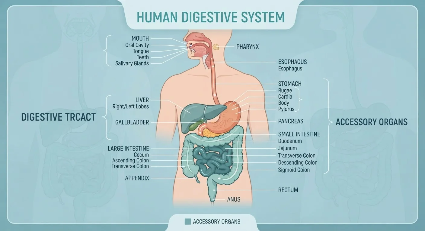
The digestive tract - also called the gastrointestinal (GI) tract - is a continuous muscular tube roughly 25 to 30 feet long that runs from the mouth to the anus. Its core job is to convert the food you eat into nutrients your body can absorb and use, while packaging up what can't be used for elimination.
This process involves four overlapping functions: ingestion (taking food in), mechanical and chemical digestion (physically breaking food apart and chemically splitting molecules), absorption (moving nutrients into the bloodstream), and elimination (expelling waste). Each organ along the tract specializes in one or more of these functions. Understanding how each segment contributes helps explain why diet, hydration, and gut health all matter - and why problems in one area often affect others. For a broader look at how digestive function intersects with other body systems, see how major body systems interact.
The Mouth and Esophagus: Where Digestion Begins
Digestion starts the moment food enters your mouth. The teeth perform mechanical digestion by cutting and grinding food into smaller pieces, increasing its surface area for chemical attack. Simultaneously, three pairs of salivary glands secrete roughly one to two liters of saliva per day. Saliva contains salivary amylase, an enzyme that begins hydrolysing (splitting) starch molecules into shorter sugar chains.
The tongue shapes chewed food into a cohesive ball called a bolus and pushes it toward the throat. Swallowing is a remarkably coordinated reflex involving more than 25 muscles. The epiglottis - a small flap of cartilage - folds over the airway to prevent food from entering the lungs.
From the throat, the bolus enters the esophagus, a muscular tube about 10 inches long. The esophagus moves food downward through rhythmic muscular contractions called peristalsis - the same wave-like mechanism that propels material through the entire GI tract. At the base of the esophagus, the lower esophageal sphincter (a ring of muscle) relaxes to allow food into the stomach, then closes to prevent stomach acid from flowing back up. When it doesn't close properly, acid reflux results.
Chewing food thoroughly - aiming for 20 to 30 chews per bite - meaningfully reduces the workload on the stomach and small intestine by increasing surface area before chemical digestion even begins.
Mechanical breakdown in the mouth is the only stage the body can consciously control, and research on gastric emptying confirms that particle size directly affects digestion rate and satiety.
Staying well-hydrated supports every stage of digestion - from saliva production to stool formation - but large volumes of water consumed immediately with a meal can dilute digestive enzymes slightly, so sipping steadily throughout the day is generally preferable.
Adequate fluid intake is consistently associated with healthy transit times and reduced risk of constipation, according to gastroenterology guidelines.
The Stomach: A Muscular Mixing Chamber
The stomach is a J-shaped, expandable muscular organ that can hold up to about one liter when comfortably full - and up to four liters at its maximum stretch. It performs three main tasks: storing food temporarily, mechanically churning it, and chemically digesting proteins.
Specialized cells lining the stomach wall secrete hydrochloric acid (HCl) and the enzyme pepsin. The acid drops the stomach's pH to between 1.5 and 3.5 - strongly acidic - which denatures (unfolds) proteins, making them accessible to pepsin for breakdown. A thick layer of mucus protects the stomach lining from digesting itself. Coordinated muscular contractions churn the food with these secretions over two to four hours, producing a semi-liquid mixture called chyme.
The stomach releases chyme in small, controlled spurts through the pyloric sphincter into the small intestine. This gating function prevents the intestine from being overwhelmed and ensures thorough mixing with digestive secretions downstream.
The Small Intestine: Where Most Absorption Happens
Despite its name, the small intestine is the longest segment of the GI tract - roughly 20 feet - and is where approximately 90% of nutrient absorption takes place. It is divided into three sections: the duodenum, the jejunum, and the ileum.
The duodenum (the first 10 inches) is where chyme mixes with digestive enzymes from the pancreas and bile from the liver, neutralising the acid and breaking fats, proteins, and carbohydrates into absorbable units. The jejunum is the primary site of nutrient absorption. The inner wall is lined with tiny finger-like projections called villi, each covered in even smaller microvilli, collectively forming the brush border. This architecture amplifies the absorptive surface area to the equivalent of a tennis court. The ileum absorbs whatever nutrients weren't captured earlier, including vitamin B12 and bile salts, which are recycled back to the liver.
Nutrients absorbed through the intestinal wall pass into capillaries and are carried to the liver via the portal vein for processing before entering general circulation.
The Large Intestine and Elimination
Material that wasn't absorbed in the small intestine - mostly water, electrolytes, fiber, and dead cells - passes into the large intestine (colon), which is wider but only about five feet long. The colon's primary job is water and electrolyte reclamation: it absorbs roughly 90% of the water remaining in the material it receives, converting liquid chyme into formed stool over 12 to 48 hours.
The large intestine also hosts the gut microbiome - trillions of bacteria, fungi, and other microorganisms. These microbes ferment dietary fiber that human enzymes can't digest, producing short-chain fatty acids that nourish colon cells and influence immune function. Research continues to map the microbiome's broader effects on health.
Formed stool collects in the rectum until stretch receptors signal the urge to defecate, at which point voluntary muscles control elimination through the anus. Transit time through the entire GI tract varies widely - typically 24 to 72 hours - influenced by diet, hydration, physical activity, and individual physiology.
Supporting Organs: Liver, Pancreas, and Gallbladder
Three organs outside the GI tube itself play indispensable roles. The liver produces bile - a fluid that emulsifies dietary fats, breaking large fat globules into smaller droplets so lipase enzymes can access them. The liver also processes absorbed nutrients, filters toxins from portal blood, and manufactures proteins needed for clotting and transport.
The gallbladder stores and concentrates bile between meals, releasing it into the duodenum when fatty food arrives. The pancreas is both an endocrine gland (producing insulin and glucagon to regulate blood sugar) and an exocrine gland, secreting a suite of digestive enzymes - lipase, proteases, and amylase - along with bicarbonate to neutralize the acid in incoming chyme.
These organs are connected to the duodenum via ducts and operate in precise coordination with hormonal signals triggered by the arrival of food. For more on how anatomy content is taught and where common misconceptions arise, see reliable anatomy education resources.
This article is for general educational purposes only and is not a substitute for professional medical advice, diagnosis, or treatment. Consult a qualified healthcare provider with questions about your digestive health.