

Key Takeaways
Cardiac Anatomy
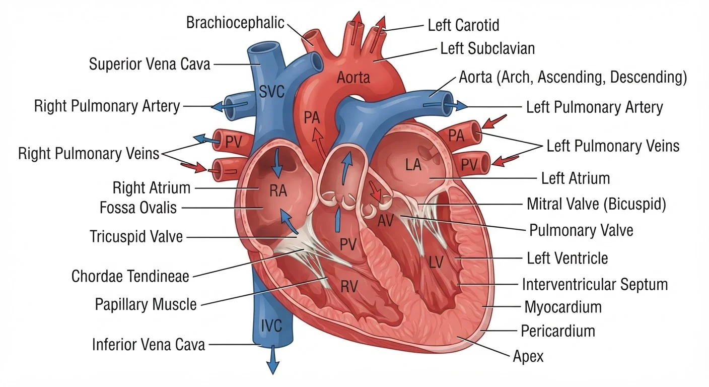
Cardiac anatomy refers to the physical structure of the heart - its chambers, walls, valves, and connected blood vessels. The human heart is a muscular, four-chambered organ roughly the size of a fist, located slightly left of center in the chest. Its design allows it to simultaneously manage two separate circulation loops: one to the lungs and one to the rest of the body.
The heart wall itself is composed of three distinct layers - the epicardium (outer), myocardium (middle muscular layer), and endocardium (inner lining) - each playing a structural or functional role in cardiac contraction.
The Heart as a Two-Pump System
Most people think of the heart as a single pump, but it is more accurately understood as two coordinated pumps sharing one muscular structure. The right side manages pulmonary circulation - sending oxygen-depleted blood to the lungs to be refreshed. The left side handles systemic circulation - distributing freshly oxygenated blood to every organ, muscle, and tissue in the body.
These two circuits run simultaneously with every heartbeat, kept separate by the septum, a thick internal wall. Understanding this dual-pump design is the key to making sense of how each chamber and valve fits into the larger picture. For a grounding in the directional language used to describe heart structures, see key anatomical terms used in human anatomy.
The Four Chambers: A Room-by-Room Walkthrough
Blood enters the heart's right atrium - the first "room" - through two large veins: the superior vena cava (carrying blood from the upper body) and the inferior vena cava (from the lower body). The right atrium acts as a receiving chamber, holding blood briefly before contracting and pushing it through the tricuspid valve into the right ventricle.
The right ventricle is a stronger, thicker chamber. When it contracts, it forces blood through the pulmonary valve and into the pulmonary artery, sending it to the lungs where carbon dioxide is exchanged for oxygen.
Oxygenated blood returns from the lungs via the pulmonary veins and arrives in the left atrium. From there, it moves through the mitral valve (also called the bicuspid valve) into the left ventricle - the heart's most powerful chamber. The left ventricle's walls are roughly three times thicker than the right ventricle's, built to generate the pressure needed to push blood through the aortic valve and into the aorta, the body's largest artery.
~5 L
Blood pumped per minute at rest
The adult heart circulates the body's entire blood volume - roughly 5 liters - approximately once per minute at a resting heart rate, according to standard cardiovascular physiology references.
100,000
Heartbeats per day
At a typical resting rate of 60-100 beats per minute, the heart completes approximately 100,000 contractions every 24 hours, according to the American Heart Association.
0.8 sec
Duration of one cardiac cycle
One complete heartbeat - including both contraction (systole) and relaxation (diastole) phases - takes approximately 0.8 seconds at a resting heart rate of 75 beats per minute.
The Valves: Gatekeepers of Blood Flow
The heart's four valves - tricuspid, pulmonary, mitral, and aortic - open and close with each heartbeat to maintain one-directional flow. They are structured from thin, flexible leaflets of tissue anchored by fibrous rings. Valves on the right side of the heart (tricuspid and pulmonary) handle lower-pressure flow; those on the left (mitral and aortic) withstand considerably higher pressures.
When valves malfunction - either leaking backward (regurgitation) or failing to open fully (stenosis) - the heart must work harder to compensate. The characteristic sounds a physician hears with a stethoscope - the classic "lub-dub" - are the sounds of the mitral and tricuspid valves closing ("lub") followed by the aortic and pulmonary valves closing ("dub").
How to Remember the Valve Names
A useful memory aid: the valves on the right side (tricuspid and pulmonary) both start with letters in the first half of the alphabet, while those on the left (mitral and aortic) come later. More practically, associate each valve with its location - the tricuspid sits between the right atrium and right ventricle, the mitral between the left atrium and left ventricle, and the semilunar valves (pulmonary and aortic) guard the exits to the great vessels.
Major Vessels: The Heart's Entry and Exit Points
Four sets of major blood vessels connect directly to the heart, forming the entry and exit points of both circulation loops:
- Superior and inferior vena cava: Return deoxygenated blood from the body to the right atrium.
- Pulmonary artery: Carries deoxygenated blood from the right ventricle to the lungs - one of the few arteries in the body that carries oxygen-poor blood.
- Pulmonary veins: Return oxygenated blood from the lungs to the left atrium - one of the few veins that carry oxygen-rich blood.
- Aorta: The largest artery in the body, distributing oxygenated blood from the left ventricle to the entire systemic circulation.
The heart also has its own blood supply delivered by the coronary arteries, which branch off the aorta just above the aortic valve. Blockages in these vessels are the underlying cause of most heart attacks.