

Key Takeaways
Start here
The Respiratory System at a Glance
Next
Upper Respiratory Tract: Where Air Enters
Then
Lower Respiratory Tract: From Trachea to Alveoli
Go deeper
How Gas Exchange Actually Works
Apply it
What Can Go Wrong - and Why It Matters
The Respiratory System at a Glance
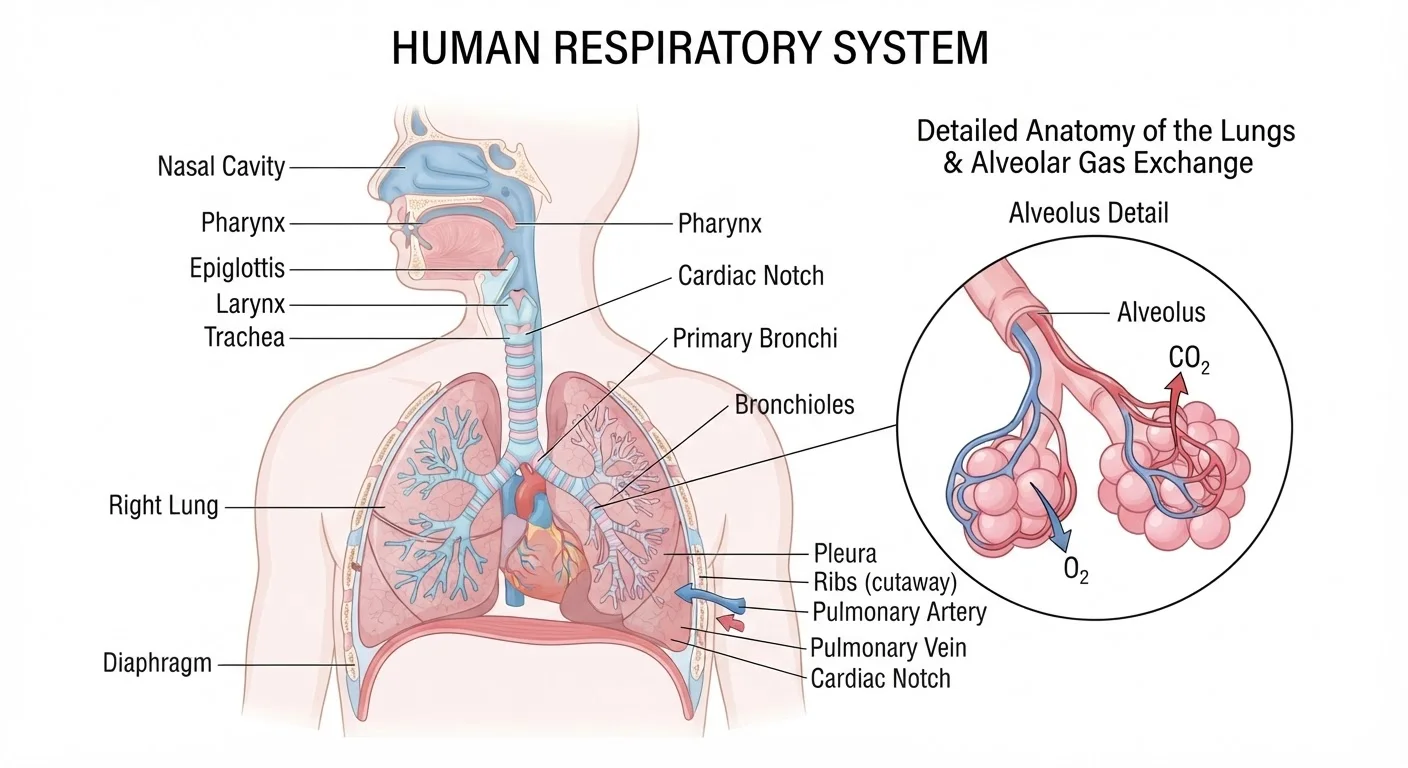
Most people think of breathing as a lung activity. In reality, each breath you take is managed by a relay team of structures - six major ones - before oxygen reaches your blood. Understanding who does what reveals why respiratory health is far more nuanced than simply keeping your lungs clear.
Physiologists divide the system into two broad zones: the upper respiratory tract (nose, pharynx, and larynx) and the lower respiratory tract (trachea, bronchi, and lungs including the alveoli). Air passes through every one of these in sequence with each inhale.
Alveoli
Microscopic air sacs at the ends of the airways inside the lungs where oxygen passes into the blood and carbon dioxide passes out.
Cilia
Tiny hair-like structures lining the airways that sweep mucus and trapped particles away from the lungs.
Diffusion
The passive movement of molecules from an area of high concentration to an area of low concentration - the mechanism driving gas exchange in the alveoli.
Bronchi
The two main airways branching off the trachea, one leading into each lung, that divide further into smaller bronchioles.
Epiglottis
A small cartilage flap in the throat that automatically closes over the airway when you swallow, preventing food and liquid from entering the lungs.
Gas exchange
The process by which oxygen moves from inhaled air into the bloodstream while carbon dioxide moves from the blood into the lungs to be exhaled.
Upper Respiratory Tract: Where Air Enters
The journey begins at the nose. The nasal cavity is lined with a moist mucous membrane and fine hairs called cilia. Together they trap airborne particles, warm cold air to near body temperature, and add humidity - protecting the delicate tissue further down.
Air then moves into the pharynx (throat), a shared corridor for both food and air. Just below sits the larynx (voice box), which houses the vocal cords and, critically, the epiglottis - a flap of cartilage that seals the airway every time you swallow, preventing food from entering the lungs.
When the larynx is irritated by a particle that slips through, the reflex cough you experience is the system's emergency override, generating airflow speeds that can exceed 500 miles per hour to eject the intruder.
Lower Respiratory Tract: From Trachea to Alveoli
Below the larynx, air enters the trachea - a rigid tube roughly 4-5 inches long reinforced by C-shaped rings of cartilage that keep it open even during forceful breathing. Its inner lining continues the mucus-and-cilia filtration started in the nose.
The trachea splits at a junction called the carina into two bronchi, one feeding each lung. Those bronchi branch repeatedly into smaller bronchioles, forming a tree-like network. The smallest bronchioles terminate in clusters of alveoli - the true workhorses of the system.
Research published in the American Journal of Respiratory and Critical Care Medicine estimates that the average adult lung contains approximately 480 million alveoli. Spread flat, their combined surface area would cover roughly 70 square meters - about the size of a singles tennis court - providing an enormous interface for gas exchange.
How Gas Exchange Actually Works
Gas exchange is governed by diffusion - molecules naturally move from areas of higher concentration to lower concentration without any energy input required. When oxygen-rich air fills the alveoli, oxygen concentration there is higher than in the surrounding capillary blood, so oxygen crosses the ultra-thin alveolar membrane into the bloodstream.
Carbon dioxide, a waste product of cellular metabolism, follows the opposite gradient: it is more concentrated in the blood than in the alveolar air, so it diffuses out into the lungs and is expelled when you exhale.
The alveolar walls are only about 0.2 micrometers thick - roughly 300 times thinner than a human hair - which is precisely what makes this passive transfer fast enough to supply the body's continuous demand for oxygen.
Breathe Through Your Nose When Possible
Nasal breathing activates the full filtration and humidification system before air reaches your lungs. Mouth breathing bypasses most of those defenses, delivering drier, less-filtered air directly to the trachea. During exercise, mouth breathing is often necessary, but returning to nasal breathing during rest helps protect the lower airways.
What Can Go Wrong - and Why It Matters
Because the respiratory system is a sequence of dependent structures, a problem in any one segment can disrupt the whole pathway. Common conditions map cleanly onto the anatomy:
- Rhinitis and sinusitis inflame the upper nasal passages, reducing filtration and airflow.
- Laryngitis swells the larynx, distorting or silencing the voice and narrowing the airway.
- Asthma causes the bronchi and bronchioles to narrow due to inflammation and muscle spasm, restricting airflow to the alveoli.
- Chronic Obstructive Pulmonary Disease (COPD) - most often caused by long-term smoking - progressively destroys alveolar walls, collapsing the gas-exchange surface area.
- Pneumonia fills alveoli with fluid or pus, blocking the diffusion process at its final stage.
Understanding where in the system a condition strikes helps clinicians target treatment precisely. It also underscores why protecting the entire respiratory pathway - not just the lungs - is central to long-term health.
This article is for general educational purposes only and is not a substitute for professional medical advice, diagnosis, or treatment. Always consult a qualified healthcare provider with any questions about a medical condition.